|
РАЗМНОЖЕНИЕ
ТРОМБОЦИТОВ
ИЛЛЮСТРАЦИИ |
PROPAGATION
OF THROMBOCYTES ILLUSTRATION |
|
|
|
|
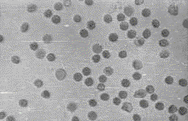
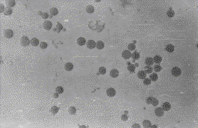
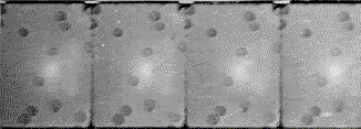
Фото
1. Нативный
препарат.
Кровь,
разведённая
3%-ным раствором
хлористого
натрия в
воде (1:200) в
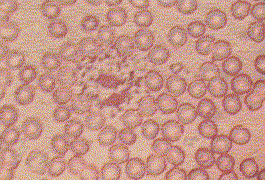
камере. Начало наблюдения. Увеличение в 500 раз. Photo 1. Native preparation. Blood diluted by 3% sodium chloride aqueous solution (1:200) in the chamber. The begining of observations. 500h magnification. |
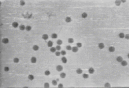
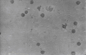
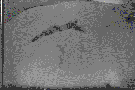
Фото
2. То же через 84
часа. В
верху
виден
овальный
тромбоцит.
Он свободный. Photo 2.
The same after 84 hours. There is free oval thrombocyte
below. |
|
|
|
|
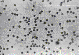
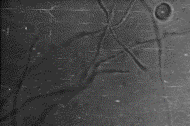
Фото
3. То же через 132
часа 5 минут
(увеличение
уменьшено). Photo 3.
The same after 132 hours and 5 minutes. |
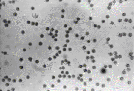
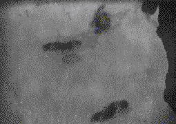
Фото
4. То же через 132
часа 5 минут 10
секунд.
Можно
оценить
движение цепочек
и отдельных
тромбоцитов
в сравнении
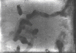
с фото 3. Photo 4. The same after 132 hours, 5 minutes and 10 seconds. One can evaluate the motion of some chains and thrombocytes. In comparison with the photo 3. |
|
|
|
|
Фото
5. То же через 216
часов.
Камера
смещена для наблюдения
за
тромбоцитом
диаметром 5
мкм. У него
хорошо
видно
изменение
формы при его
колебаниях. Photo 5. 1The same after 216 hours . The same is moved in ordercto observe the thrombocyte
with diameter 5 mkm. The changes
of the shape is visible during oscillations of the thrombocyte. |
Фото
6. То же через 336
часов. Photo 6.
The same after 336 hours. |
|
|
|
|
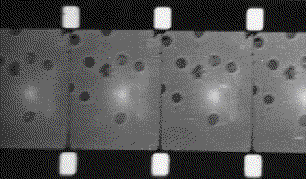
Фото
7. Кровь,
разведённая
3%-ным водным
раствором
хлористого
натрия (1:200).
Увеличение
в 400 раз скорость
съёмки 24
кадра в
секунду.
Правее
центра кадра
виден
эритроцит,
сильно
деформированный,
который
энергично
двигается,
иногда перекатывается.
От него в
процессе
наблюдения
было
замечено
отделение
трёх тромбоцитов
в виде
палочек,
после чего
его форма
стала
сравнимой с
формой
остальных
эритроцитов.
Photo 7.
Blood diluted by 3% sodium chloride aqueous solution (1:200). 400 magnification, speed of filming is frames per second.
There is very deformed erythrocyte on the right of the centre
which moves actively and roll sometimes. Separation
of 3 bacillus-shaped thrombocytes were
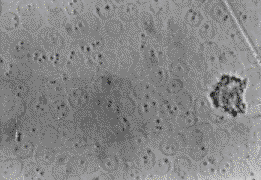
observed. After that its shape became comparable with the other erythrocytes. |
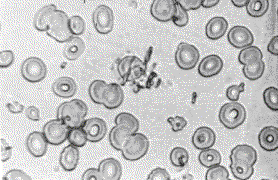
Фото
8. Мазок крови
больной
септическим
эндокардитом
в острой
фазе.
Фиксация
смесью
Никифорова.
В центре
ясно виден
тромбоцит,
прикреплённый
к
эритроциту. Photo 8. Smear of blood of patient sick with septic endocardit, in acute phase. There are thrombocytes. |
|
|
|
|
Фото 9.
Тот же
препарат,
что и на фото 7,
через 4 часа
после
приготовления.
В средней
части кадра
виден
тромбоцит в
виде слегка
изогнутой
палочки
длиной 22 мкм
движущейся,
изгибаясь (24
кадра в
секунду) Photo 9.
The same preparation as on the Photo 7, in 4 hours after preparing. There is thrombocyte in the middle part looking like a little
curved bacillus with a length of 22 mkm. It moves
sinuously. Speed of filming is 24 frames pro second. |
Фото 10.
Тромбоциты
были отмыты,
помещены на формваровую
плёнку и контрастированы
напылением
тонкого
слоя
платины под
углом 70 градусов. увеличение
электронного
микроскопа 5000
раз. Цепочка
состоит из
тромбоцитов
разных размеров
во втором
(справа )
тромбоците
определяется
почка с
правого конца.
Виден выход
почки из
второго
тромбоцита.
У последнего
видны
тонкие
выросты,
заканчивающиеся
расширением.
Заметна ассиметрия полюсов клеток. Тромбоциты культивировались. Photo
10. The thrombocytes were washed, placed on the
film for electron microscopy and contrasted by spraying of the thin platinum
layer at the angle of 70 grad .Electron microscope has 5000x magnification. The
chin consist of the thrombocytes
of different sizes. There is a growing gemma at the
right side of the third right thrombocyte. One can
see the going out of the gemma from second thrombocyte. The last thrombocyte
has the thin projections on the ends. The poles of the cells are asymmetric.
The thrombocytes were cultivated. |
|
|
|
|
Фото 11.
Кровь
посеяна на
стерилизованный
фильтрованием
жидкий
гематоген.
Видны длинные
нити -
колонии
тромбоцитов,
которые двигались.
Нити не
распластаны
по дну
камеры. Photo
11. Sowing of blood on the liquid hematogen
sterilized by filtration. There are the long threads - the colonies of thrombocytes which are moving. The thread
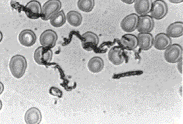
are not spread on the bottom of the chamber. |
Фото 12.
См. фото 10 -
другой
участок
препарата. В нижней
части
снимка
видна
слегка
изогнутая
палочка, в которой
произошло
разделение
ядер, но не
закончилась
перешнуровка.
Photo
12. See photo. Here is another part of preparation. There is a little curved
bacillus on the photograph. The division of the nuclei is finished but the construction still continue. |
|
|
|
|
Фото 13.
Другой
участок
препарата,
снятого на
фото 10. Photo
13. Another fragment of the preparation from the photo 10. |
Фото 14.
Мазок крови
больной
септическим
эндокардитом
в период
лихорадки. Photo
14. Smear of blood of the patient sick with septic endocardit
in fever phase. |
|
|
|
|
Фото 15.
Мазок крови
Фиксация
парами
дихлорэтана.
Photo
15. Fixation by vapours of dichlorethane. |
Фото 16.
Свищ,
образовавшийся
на средней трети
правого
бедра у
больной
хроническим
септическим
эндокардитом
(глубина
верхнего
края камеры 15
мм, объём 30 куб
см). из
содержимого
свища были
выделены
только тромбоциты.
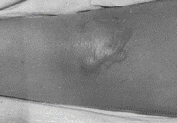
Photo 16. Fistula on the middle third of the thigh of the patient sick with chronic septic endocardit. The depth of the chamber is 15 mm, it volume is 30 cm. Only thrombocytes were obtained from it |
|
|
|
|
Фото
17.Свежеприготовленный
мазок крови
культивировался
при 100%
влажности,
при 37 град. С, 4 - 8 часов,
затем
высушивался,
фиксировался
нагреванием
и
окрашивался.
Видны
расползшиеся
из
лейкоцита
тромбоциты. Photo 17. Fresh the blood smear was cultivated at 100 % of humidity, at 37 beg. C., 4 - 8 hours, then was dryed up, was fixed by heating and was imbued. The thrombocytes are visible crawling from a leucocyte. |
|
